 Products Home / Optical Systems / Objective Lenses, Scan Lenses, and Tube Lenses / Miniature Finite Conjugate Objectives and Scan Lenses
Miniature Finite Conjugate Objectives and Scan Lenses
- Ideal for Two-Photon Microscopy, GCaMP Imaging, and Functional Imaging
- Optimized for Imaging through Thin Windows, Prisms, and GRIN Lenses
- Air and Water Immersion Objective Options
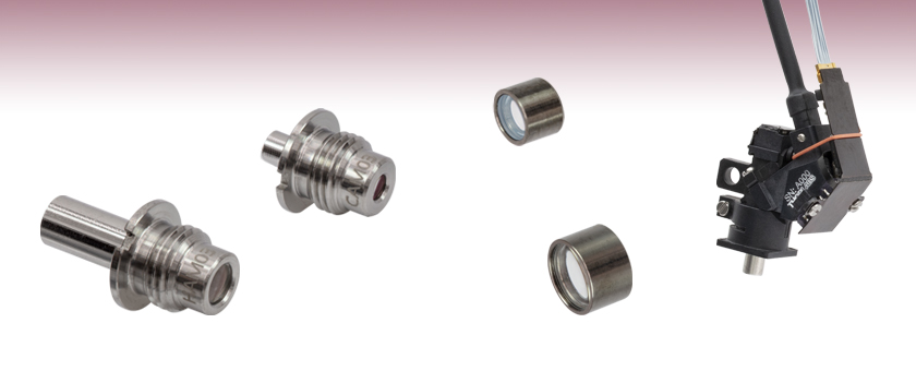
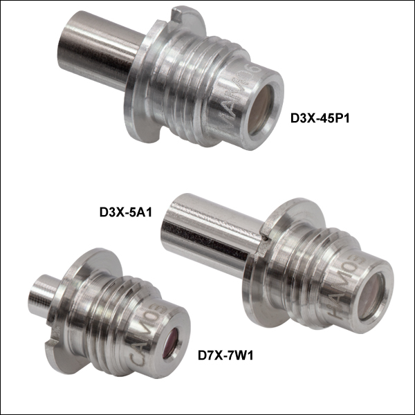
D3X-5A1
3X Miniature Objective, 0.50 NA
D7X-7W1
7X Miniature Objective,
0.70 NA
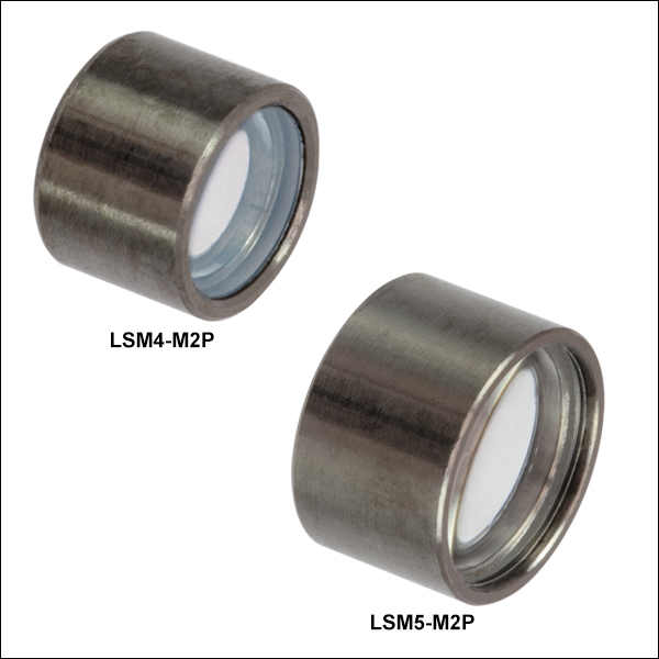
LSM4-M2P
Miniature Scan Lens,
EFL = 4.07 mm
LSM5-M2P
Miniature Scan Lens, EFL = 5.24 mm
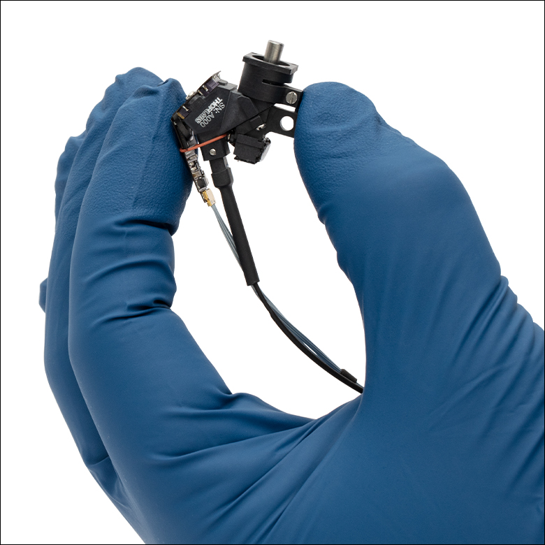
Mini2P Miniature Two-Photon Microscope with a D3X-45P1 Objective
(Not to Scale)


Please Wait
cortex expressing GCaMP6 and was taken with the Thorlabs’ Mini2P Microscope and a D3X-45P1 miniature objective. (Sample preparation courtesy of Mario Fernandez de la Puebla. Imaging performed at Dr. Weijian Zong’s lab at the Kavli Institute for Systems Neuroscience, Norway.)
Click to Enlarge
Figure 1.1 The D3X-45P1 miniature objective used with our Mini2P Two-Photon Imaging System.
Features
- Miniature Finite Conjugate Objectives and Scan Lenses
- Interchangeable Objectives for Various Applications:
- Optimized for Imaging Through Prisms, GRIN Lenses, or Thin Windows
- 3X and 7X Magnifications
- Antireflective (AR) Coatings Across the Visible and Near-IR Wavelength Ranges
- Numerical Apertures (NA) Up to 0.70
- Ideal for Two-Photon Microscopy, GCaMP Imaging, and Functional Imaging Applications
Thorlabs offers Miniature Finite Conjugate Objectives and Scan Lenses that are designed for use in miniature two-photon microscopes, such as our Mini2P Two-Photon Imaging System. These compact objectives and scan lenses are AR-coated for lower reflectance at wavelengths in the visible and near-infrared commonly used in two-photon microscopy.
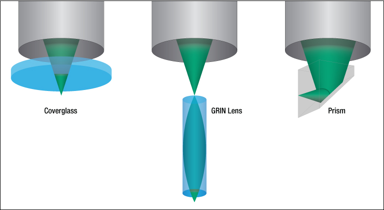
Each miniature objective is equipped with M5 x 0.5 threading, enabling interchangeability based on the user’s application. Designed for optimal imaging through prisms, GRIN lenses, or thin windows (See Figure 1.3), these objectives support imaging at various tissue depths and orientations. However, their use is not limited to these applications, offering flexibility for a wide range of techniques. The finite conjugate objectives can be used together with miniature scan lenses for two-photon microscopy, GCaMP imaging, and functional imaging applications. Video 1.2 shows the spontaneous signal of a mouse somatosensory cortex expressing GCaMP6 and was taken with the Thorlabs’ Mini2P Microscope and a D3X-45P1 miniature objective. Please see the Specs tab for additional details on the specifications of these miniature objectives and scan lenses, and the Application Info tab for more information on how these values are defined.
Click to Enlarge
Figure 1.3 The miniature objectives are optimized for imaging through prisms, GRIN lenses, and thin windows. Figure reproduced from Large-Scale Two-Photon Calcium Imaging in Freely Moving Mice.1
Click to Enlarge
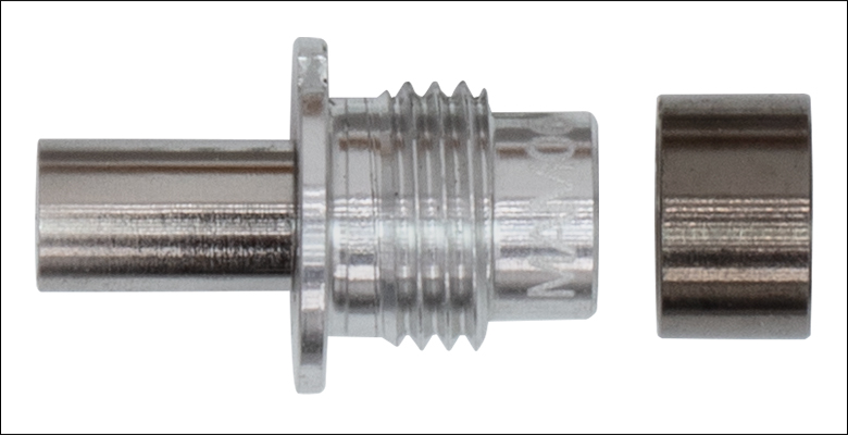
Figure 1.4 A D3X-45P1 miniature objective and an LSM5-M2P scan lens.

References
- Zong, W. et al., "Large-scale two-photon calcium imaging in freely moving mice", Cell Vol. 185, Issue 7, 1240-1256 (2022)
| Objective Specificationsa | ||||||||||||||||
|---|---|---|---|---|---|---|---|---|---|---|---|---|---|---|---|---|
| Item # | D3X-45P1 | D3X-5A1 | D3X-5W1 | D7X-7W1 | ||||||||||||
| Suggested Use Caseb | Prism | GRIN Lens | Thin Window | Thin Window | ||||||||||||
| AR Coating Wavelength Range | Ravg < 0.5% ( Ravg < 1% (89 Ravg < 2% ( |
Ravg <1% ( |
||||||||||||||
| Magnification | 3X | 3X | 3X | 7X | ||||||||||||
| Numerical Aperture | 0.45 | 0.50 | 0.50 | 0.70 | ||||||||||||
| Effective Focal Length | 9.49 mm | 10.85 mm | 8.81 mm | |||||||||||||
| Working Distancec | 2.32 mm (0.12 mm Air + 2.00 mm Glassd + 0.20 mm Water) | 0.58 mm (Air) | 1.17 mm (1.00 mm Water + 0.17 mm Coverglass) | 0.41 mm (0.24 mm Water + 0.17 mm Coverglass) | ||||||||||||
| Image Distance | 0.792 mm | 0.813 mm | 0.813 mm | 0.870 mm | ||||||||||||
| Parfocal Length | 7.50 mm | 7.16 mm | 6.77 mm | 3.36 mm | ||||||||||||
| Entrance Pupil Diameter | 7.68 mm | 8.52 mm | 8.12 mm | 8.45 mm | ||||||||||||
| Entrance Pupil Positione | 25.5 mm | 25.2 mm | 24.2 mm | 42.9 mm | ||||||||||||
| Threading | M5 x 0.5 | |||||||||||||||
| Weight | 0.40 g | 0.64 g | 0.57 g | 0.45 g | ||||||||||||
| Scan Lens Specificationsa | ||
|---|---|---|
| Item # | LSM4-M2P | LSM5-M2P |
| AR Coating Wavelength Range | Ravg ≤ 1% (900 - 960 nm & 1000 - 1060 nm) | Ravg ≤ 1% (530 - 1064 nm) |
| Clear Aperture | Ø2.5 mm | Ø3.9 mm |
| Effective Focal Length | 4.07 mm | 5.24 mm |
| Scanning Distance | 3.11 mm | 4.70 mm |
| Lens Working Distance | 3.26 mm | 4.14 mm |
| Entrance Pupil Diameter | 0.81 mm | 1.75 mm |
| Diffraction Limited Scan Angle | 11° | 2.8° |
| F-Theta Distortion | 1.90% | 0.51% |
| Axial Colorb | <36.1 µm | <46.5 µm |
| Field Curvatureb | <500 µm | <500 µm |
| Outer Diameter | Ø3.1 mm (Ø0.12") | Ø4.5 mm (Ø0.18") |
| Weight | 0.12 g | 0.21 g |
- Zong, W. et al., Large-scale two-photon calcium imaging in freely moving mice, Cell Vol. 185, Issue 7, 1240-1256 (2022)
Scan lenses are utilized in a variety of laser imaging systems, including confocal laser scanning microscopy and multiphoton imaging systems. In these applications, a laser is precisely scanned across the sample to provide point-by-point illumination, enabling the reconstruction of high-resolution 2D and 3D images. One of the functions of a scan lens in a laser scanning microscope is to project the scanning mirror to the entrance pupil of the objective such that it effectively scans at the objective entrance pupil. The system described below utilizes a miniature scan lens and a finite conjugated miniature objective. The scanning mirror, defined as the entrance pupil of the scan lens in Figure 3.1, is located to the left of the scan lens. The scan lens projects the laser input beam originating from the entrance pupil and focuses the rays on an intermediary plane to the right, defined as the image plane. The separation between the scan lens and the objective should be set such that the image plane of the scan lens coincides with that of the objective. The beam projected from the scanning mirror onto the image plane acts as the laser input for the objective, and the objective focuses this beam onto the sample plane to the right. The distance from the front element of the objective to the sample plane is defined as the working distance of the objective.
If the beams are extended from the image plane, they converge on a plane to the right—designated as the exit pupil of the scan lens and labeled in Figure 3.1 as the Virtual Aperture. Similarly, for the objective, its entrance pupil is virtually located outside the physical lens body and is also labeled as the Virtual Aperture. The position of the virtual aperture relative to the image plane is defined as the entrance pupil position of the objective, and the diameter of the virtual aperture is defined as the entrance pupil diameter of the objective (labelled as the Entrance Pupil Diameter in Figure 3.1). It is important to note that the objective's entrance pupil position and diameter can be much larger than the physical size of the objective. A laser scanning system design should ensure that the exit pupil position of the scan lens matches the entrance pupil position of the objective to achieve an optical field of view (FOV) with uniform illumination. For this to be possible, the scanning mirror must be offset from the front focal plane of the scan lens, causing the beams extending from the image plane to converge at a finite virtual position.
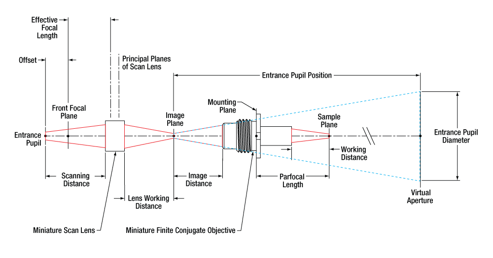
Figure 3.1 Visual Definitions of the Terms Described in Table 3.2 (Not to Scale)
| Table 3.2 Glossary of Terms | |
|---|---|
| Numerical Aperture (NA) | Numerical aperture, a measure of the acceptance angle of an objective, is a dimensionless quantity. It is commonly expressed as: NA = ni × sinθa where θa is the maximum acceptance half-angle of the objective, and ni is the index of refraction of the immersion medium. This medium is typically air, but may also be water, oil, or other substances. |
| Working Distance (WD) |
The working distance is the distance between the front element of the objective and the top of the specimen. |
| Lens Working Distance (WD) |
For scan lenses, the working distance is the distance between the tip of the scan lens housing and the back focal plane, or image plane, of the scan lens. |
| Parfocal Length | The Parfocal Length is the distance from the objective mounting plane to the sample plane. |
| Entrance Pupil Diameter (EP) | The entrance pupil diameter (EP), sometimes referred to as the entrance aperture diameter, corresponds to the appropriate beam diameter one should use to allow the objective or scan lens to function properly. For miniature scan lenses, the EP is expressed as: EP = 2 × NA × Effective Focal Length For finite conjugate miniature objectives, the EP is defined as the diameter of the beam at the virtual aperture of the objective. |
| Entrance Pupil Position | The entrance pupil position is the distance from the intermediary image plane between the scan lens and objective to the virtual aperture. |
| Image Distance | The image distance is the distance from the intermediary image plane between the scan lens and objective to the tip of the objective housing. |
| Scanning Distance (SD) | The scanning distance is the distance between the entrance pupil and the front plane of the scan lens (the left side in Figure 3.1). |
| Scan Angle (SA) |
After being routed from the scanning mirror, the laser beam is incident on the lens at an angle. This angle, measured with respect to the optical axis of the lens, is the scan angle. When listed in the specification table, it indicates the maximum allowed scan angle. |
| Axial Color |
Axial color describes the shift in the image plane across the operating wavelength range of the scan lens. |
| Field Curvature |
Field curvature describes the curvature of an objective or a scan lens' plane of focus. |
| Posted Comments: | |
| No Comments Posted |

 Zoom
Zoom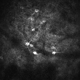
Click to Enlarge
Figure G1.2 A 350 μm FOV image of a mouse retrosplenial cortex captured by the D3X-45P1 miniature objective used with our Mini2P Two-Photon Imaging System.
Click to Enlarge
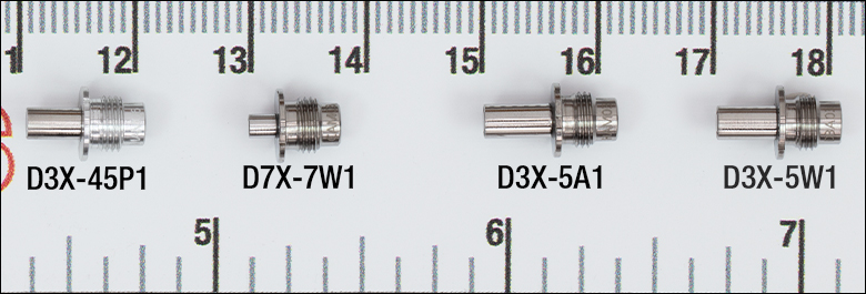
Figure G1.1 Miniature objectives are overlayed on a ruler, with major gridlines "5" and "6" separated by 1 inch. Major gridlines "15" and "16" are separated by 1 cm.
- Miniature Finite Conjugate Objectives
- Objectives Designed for Two-Photon Microscopy
- Optimized for Imaging Through Prisms, GRIN Lenses, or Thin Windows
- M5 x 0.5 Threading
Thorlabs offers compact Miniature Objectives that are designed for use with miniature two-photon microscopes, such as Thorlabs' Mini2P Two-Photon Imaging System. These objectives are optimized for imaging through prisms, GRIN lenses, or thin windows, allowing imaging to be performed at various tissue depths and orientations (See Table G1.3 for suggested use cases). The low reflectance at visible and near-infrared wavelengths commonly used in two-photon microscopy allow for a range of custom applications. For example, the D3X-45P1 objective features a longer working distance of 2.32 mm, making it well-suited for use with a prism, while also offering versatility for other applications that require extended working distances.
| Table G1.3 Key Specificationsa | ||||||||
|---|---|---|---|---|---|---|---|---|
| Item # | Suggested Use Caseb | AR Coating Wavelength Range |
Magnification | Numerical Aperture |
Effective Focal Length | Working Distancec | Threading | Weight |
| Prism | Ravg < 0.5% ( Ravg < 1% (89 Ravg < 2% ( |
3X | 0.45 | 9.49 mm | 2.32 mmd | M5 x 0.5 | 0.40 g | |
| GRIN Lens | 3X | 0.50 | 10.85 mm | 0.58 mm (Air) | 0.64 g | |||
| Thin Window | Ravg <1% ( |
3X | 0.50 | 8.81 mm | 1.17 mme | 0.57 g | ||
| Thin Window | 7X | 0.70 | 6.53 mm | 0.41 mmf | 0.45 g | |||
- Zong, W. et al., Large-scale two-photon calcium imaging in freely moving mice, Cell Vol. 185, Issue 7, 1240-1256 (2022)

 Zoom
Zoom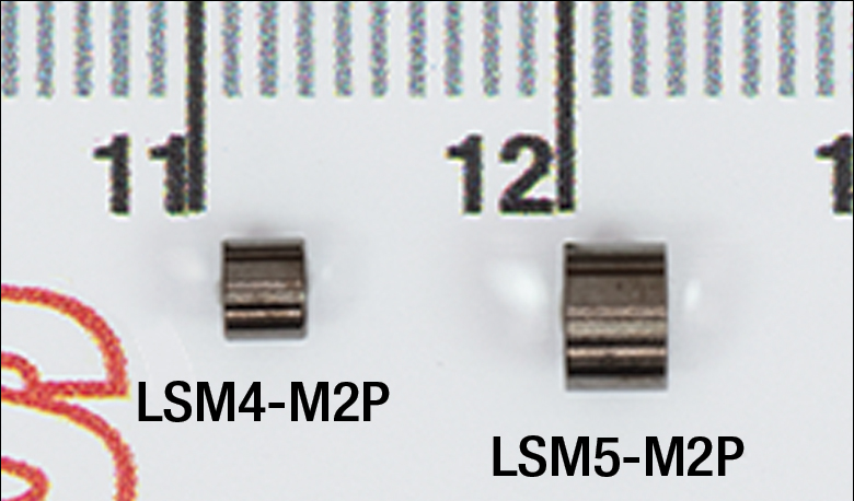
Click to Enlarge
Figure G2.2 Miniature scan lenses are overlayed on a ruler, with major gridlines "11" and "12" separated by 1 cm.
- Optimized for Two-Photon Microscopy
- LSM4-M2P: Effective Focal Length 4.07 mm, Entrance Pupil Diameter 0.81 mm, Weight 0.12 g
- LSM5-M2P: Effective Focal Length 5.24 mm, Entrance Pupil Diameter 1.75 mm, Weight 0.21 g
These miniature scan lenses are designed to work with miniature objectives for imaging tissue at different depths and orientations.
These scan lenses have AR coatings over wavelengths in the visible and NIR wavelength ranges, making them ideal for two-photon microscopy. The key specifications for these scan lenses are listed in Table G2.1. For more information on how these values are defined, see the Application Info tab.
| Table G2.1 Key Specificationsa | ||||
|---|---|---|---|---|
| Item # | LSM4-M2P | LSM5-M2P | ||
| AR Coating Wavelength Range | Ravg ≤ 1% (900 - 960 nm & 1000 - 1060 nm) | Ravg ≤ 1% (530 - 1064 nm) | ||
| Clear Aperture | Ø2.5 mm | Ø3.9 mm | ||
| Effective Focal Length | 4.07 mm | 5.24 mm | ||
| Scanning Distance | 3.11 mm | 4.70 mm | ||
| Lens Working Distance | 3.26 mm | 4.14 mm | ||
| Diffraction Limited Scan Angle | 11° | 2.8° | ||
| Outer Diameter | Ø3.1 mm (Ø0.12") | Ø4.5 mm (Ø0.18") | ||
| Weight | 0.12 g | 0.21 g | ||